
Creative Spotlight: Jewellery Inspired By Neuroscience, by Luke Maninov Hammond of Queensland Brain Institute
First published June 2, 2017
You’d be amazed by the treasures you find on Instagram.
If art and design can tell stories to engage a captive audience beyond science and medicine, Luke Maninov Hammond has done a stellar job.
I came across a snapshot of Luke’s designs via the Queensland Brain Institute’s Instagram. Luke’s work with fluorescence microscopy at the QBI has inspired his incredible jewellery designs and prints, which he has exhibited recently at Pieces of Eight gallery in Melbourne and will soon be showing at Brisbane’s Artisan Gallery from June 15.
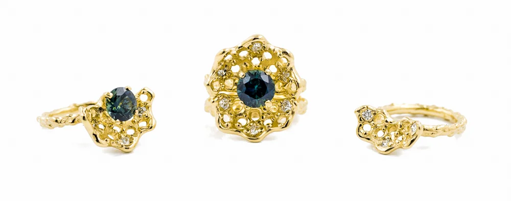
Beneath The Surface engagement ring. Luke: “This signature ring simultaneously represents the invisible worlds of cellular machinery, marine life, and dynamic connectivity within the brain. Designed as a pair, this engagement ring holds a 1.6ct Australian parti sapphire in 18ct yellow gold, surrounded by three brilliant white diamonds. Combined, the pair form a completed circle surrounding the sapphire with a golden halo set with six diamonds.” Image copyright Luke Maninov Hammond
As well as at his exhibitions, some of Luke’s work can be purchased at beneaththesurfaceprints.bigcartel.com, with profits going towards schizophrenia research. He’s also at www.lukemaninov.com, and on Instagram@lukemaninov and Facebook www.facebook.com/lukemaninovjewellery. Luke kindly provided insights for us about the mix of art, design and neuroscience while preparing for a new role at Columbia University, New York City.
Can you tell us about your neuroscience career?
When I was studying neuroscience, all I wanted to do was get involved in research on consciousness, but I took a side step and started an imaging project in a cancer biology lab with Prof. Jennifer Stow at the University of Queensland. This is when I first started using microscopes to image fluorescent proteins in cells.
It’s one thing to know that without your awareness, every single cell in your body is almost vibrating with activity and something entirely different to actually see it with your own eyes. Looking down into an ocean of darkness and seeing dynamic glowing worlds alive within cells was a profound experience that completely captured my imagination.
A few years later I had the opportunity to join the Queensland Brain Institute (QBI), where I worked to establish an imaging facility so these techniques could be used to study the brain. It’s been an incredible journey to see fluorescent imaging move from allowing us to see inside single cells, to watching neurons within the brain flashing with activity. After almost 9 years with QBI I’m about to start a new position managing and establishing a new facility at the Zuckerman Mind Brain Behaviour Institute, Columbia University in New York City.
What did your work at QBI focus on?
My work at QBI was focused on establishing a microscopy facility that offered the world’s most advanced imaging capabilities and working with QBI’s researchers to ensure they could use these instruments to make novel discoveries about the brain. This included working on projects trying to understand how neurotransmitters are released, how axons regenerate, and how to treat diseases like Alzheimer’s or motor neurone disease.
Most recently my colleagues and I published a new paper in Molecular Neurobiology which contributes key insights into how vitamin D deficiency during embryonic development can alter the brain’s dopamine system.
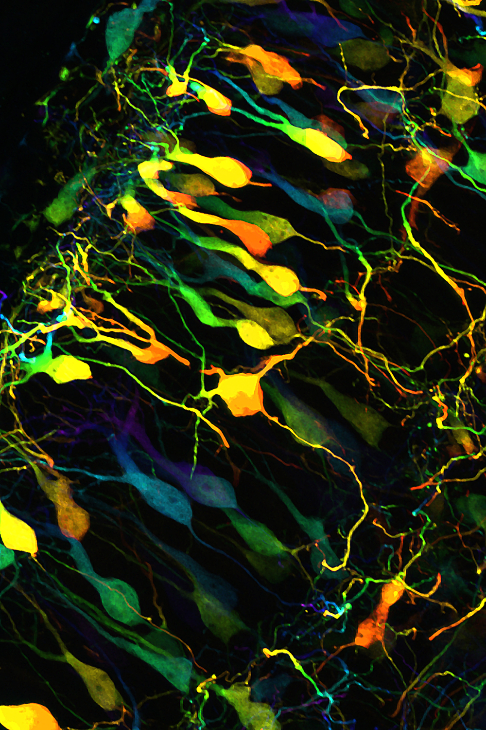
Luke: “Within the In-Between” reveals the brain cells and their complex interwoven processes. To create this image, varying colours have been used to reflect the changing depths of the neuronal processes as they extend through the brain. This image was captured at high-resolution in 3D using state-of-the-art fluorescence microscopy at the University of Queensland’s Queensland Brain Institute.” This is one of several limited-edition prints that can be purchased for schizophrenia research. Image copyright Luke Maninov Hammond
Fluorescence microscopy essentially makes the invisible worlds within cells and brains visible with glowing proteins and dyes.
Due to recent advancements we can now see objects down to 20nm in size, that’s 5000x finer than a human hair, in living cells and tissue. It’s truly amazing what we can achieve. We are in the midst of a revolution for biomedical imaging, it’s a very exciting time for brain science.
How did you start making jewellery and fine art?
I started making jewellery and objects as a new creative outlet and a way of exploring 3D form. I primarily use a technique called “Lost-Wax Casting,” which involves sculpting and creating a wax object that can be transferred into precious metal.
I fell in love with the analogue process of working with my hands to create these forms, and have been experimenting with it ever since. There is an inherent joy in creating something out of nothing based on an idea which emerges at the edge of your imagination.
Agreed! What was the point when you realised the link between your neuroscience work and a physical expression of creativity?
My practice has always been about reimagining biological form to explore themes of impermanence, consciousness and connection between living things. From the beginning I think the jewellery I was creating was informed by the 3D imaging and analysis I was performing in science but a few years in I realised it made sense to explore neuroscience more deliberately.
What, if any, resistance or challenges have you had to overcome from others, or self-doubts from yourself, when crossing between the science and fashion/design worlds?
Certainly I’ve learnt to overcome a lot of self-doubt in teaching myself to create jewellery. Navigating a path between science and art can be challenging too, in the sense that you don’t want your involvement in one role to call into question your capacity for the other. While there can be a lot of overlap, especially in creativity and coming up with ideas, in science we are required to make unbiased, precise and accurate measurements in order to understand complex processes, and this is not always the work of an artist.
On the other hand, we are exploring an unseen world for the first time, and there is an important role for art to play in sharing this with rest of the world and communicating these discoveries in ways that capture our imagination. It’s encouraging to see growing interest in bridging the worlds of art and science.
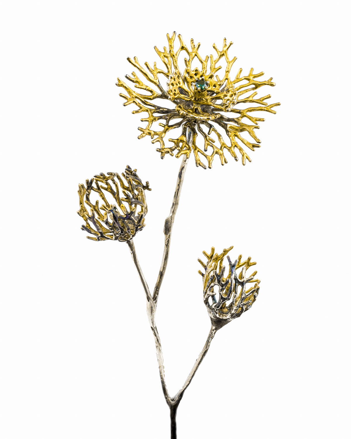
Unfolding Object. Sterling silver, Australian sapphires, gold vermeil and patina. Housed in glass bell jar. Image copyright Luke Maninov Hammond
How did the Pieces of Eight exhibition come about (in Melbourne)?
Melanie Katsalidis, the Director of Pieces of Eight, began representing my work last year. When the possibility to propose an exhibition came up, I put forward the concept of “Beneath the Surface.” I only had a few weeks to work on the show, but the timing ended up perfect as I was able to complete the project just in time to be ready to move to New York.
What do you hope people will learn or gain from your exhibition?
The exhibition explores the story of green fluorescent protein, the glowing protein discovered in jellyfish by Osamu Shimomura, from which fluorescence microscopy and our ability to see the invisible stems. It draws parallels between the unfolding microscopic structures beneath our skin and those in the depths of the ocean.
I think it’s a story stranger than fiction, that our exploration of the sea has enabled us to illuminate the living brain and journey inwards.
I hope people will come away with an interest in what is being discovered in neuroscience and a sense of wonder in the hidden beauty within us. Part of the exhibition includes large-format cellular images of the brain, which we rarely get to share with the public, so I hope this will capture people’s imaginations.
As with my other work, the pieces represent the unfolding nature of life, encouraging reflection on our coming out of the world, rather than coming into it.

“Enclosed Radial” ring. Sterling silver, Australian sapphires, gold vermeil, patina. Image copyright Luke Maninov Hammond
Neuroscience and ageing can be very intimidating subjects; how can we make the brain and neuroscience more accessible to others?
It’s true, people can put a mental block on understanding topics like this as they appear intimidating. This is why it’s important for science to engage with artists and communicators to come up with novel ways of sharing discoveries and breaking down the barrier that exists between science and the general public.
I think microscopy has a key role to play here too, the images we capture are able to directly convey the story of disease and how the brain works. Often these images are never seen by more than one or two people, as they are distilled into graphs and data points for publication, but I hope we can find more ways of sharing them more broadly. I’ve seen some amazing reactions to the few images I’ve been able to share in my exhibitions and believe they hold a capacity to spark a genuine interest in science and self discovery.
Thank you Luke for your inspiration! Don’t forget to visit his “Beneath the Surface” exhibition at Artisan Gallery, Brisbane from June 15 and buy his prints at beneaththesurfaceprints.bigcartel.com. Profits from sales go towards schizophrenia research.